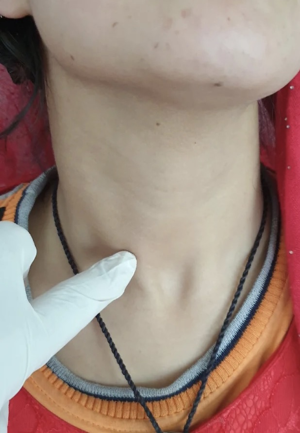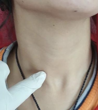
.یک خانم 22 ساله از کتله در قدام عنق یا غده درقیه شاکیست
معاینه با سوزن باریک فولیکولر نیوپلازم را نشان میدهد که میتواند ادینوم یا هم کارسینوم باشد و فرق بین این دو در بیوپسی آشکار میشود. در بیوپسی چون تهاجم کپسول تومور و اوعیه های تومور توسط تومور دیده نشد پس این یک ادینوم بوده نه کارسینوم (جهت معلومات بیشتر متن انگلیسی و تصاویر ذیل را مرور نمایید)
- Follicular Adenoma: Benign tumor that shows evidence of follicular differentiation but lacks evidence of capsular and vascular invasion and lacks papillary carcinoma nuclear features.
- Epidemiology: Adults, usually 20 – 50 years , M:F = 1:6
Clinical features / diagnosis:
- Presents with long standing solitary thyroid nodule
- Almost always solitary; if multiple, diagnose as multinodular goiter with adenomatous change.
Laboratory: Patient is usually euthyroid
Radiology description: Usually “cold” nodule, may be “warm” but rarely “hot”
Treatment: Lobectomy (not enucleation)
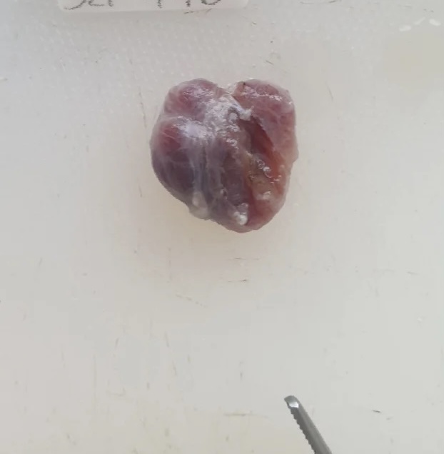
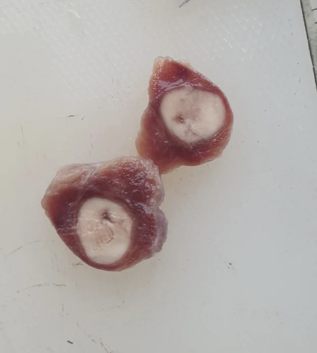
Gross description:
- Solitary, encapsulated, variable size (1 – 10 cm), In our case tumor size was 1.5 cm.
- Solid, fleshy, tan to light brown
- Bulges when fresh, compresses adjacent thyroid
- Resembles multinodular goiter due to secondary changes of hemorrhage and cystic degeneration
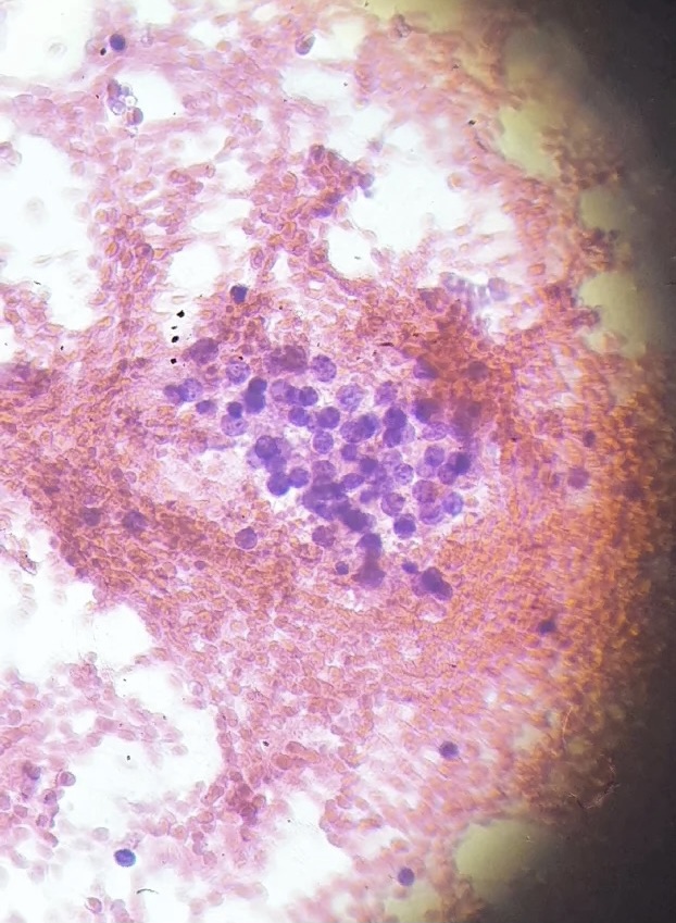
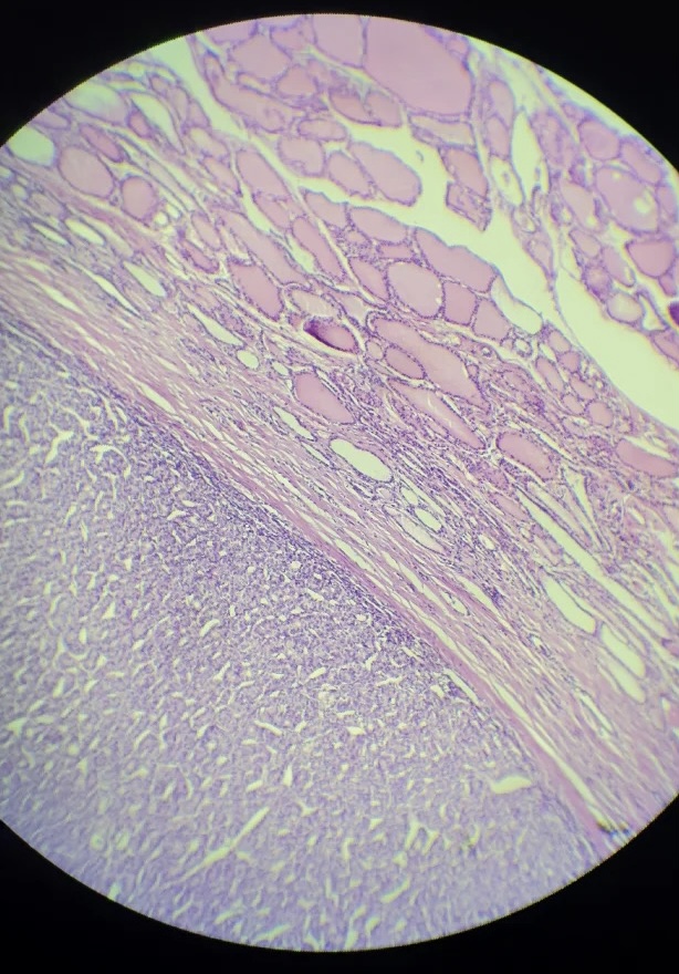
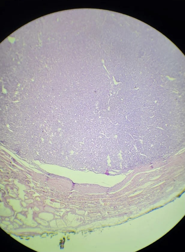
Microscopic (histologic) description:
- Completely enveloped by thin fibrous capsule.
- Architecturally and cytologically different from surrounding gland; surrounding thyroid tissue shows signs of compression
- Closely packed follicles, trabeculae or solid sheets
- Cuboidal to low columnar cells, pale staining with round inconspicuous nucleoli
- Mitoses are uncommon
- Commonly secondary changes of hemorrhage, hemosiderin deposition, sclerosis, edema, necrosis and cystic changes
- No capsular or vascular invasion after thorough sampling (at least 10 blocks), no / rare mitotic figures, no papillary nuclear features
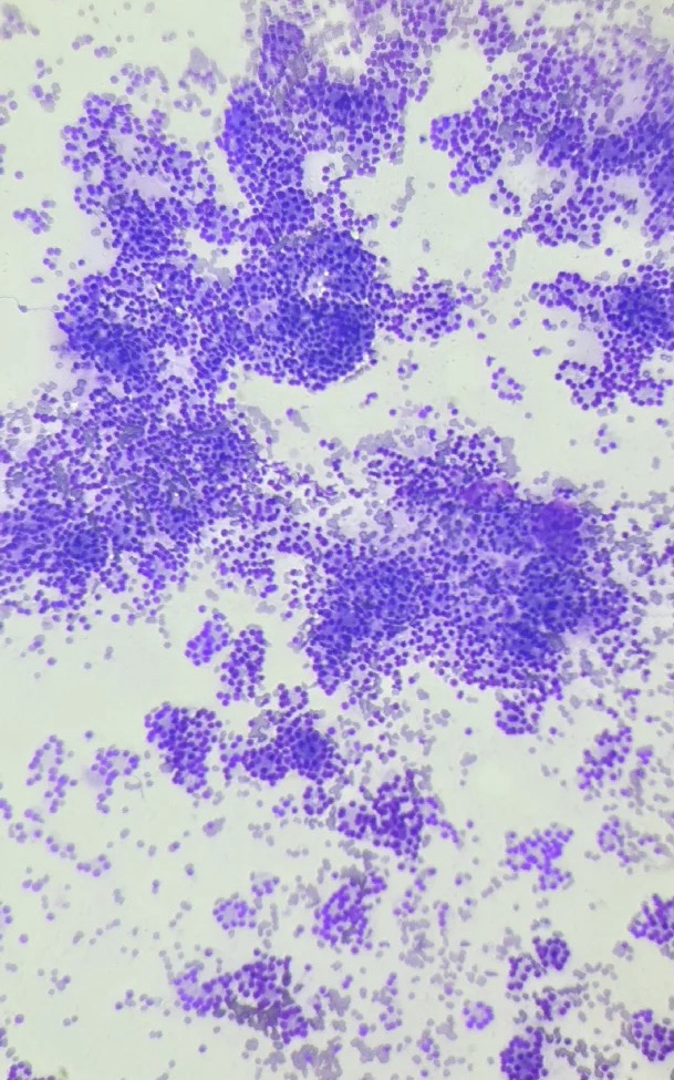
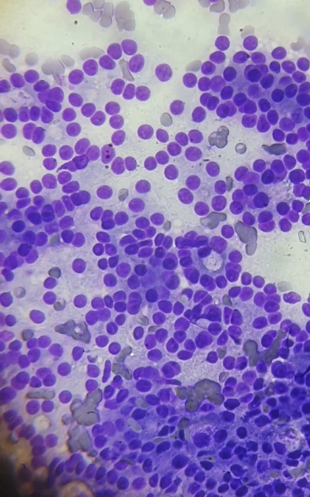
Cytology description:
- High cellularity, syncytial 3 dimensional arrangement, prominent nuclear crowding with some follicular arrangemen, but no papillary nuclear features, minimal colloid.
- Cannot rule out carcinoma based on cytologic findings.